Combined/hybrid systems literature
Downloads: Raman-SPM/AFM
-
 Product note: inVia™ confocal Raman microscope and Dimension AFM integration
The integration of the inVia confocal Raman microscope with the Dimension series AFMs combines industry leading instruments to provide a solution that gives the best possible performance:
-
 Product note: Renishaw Raman-AFM/TERS solutions
Product note - Combined Raman/AFM (atomic force microscope) systems are excellent for characterising the properties of materials at sub-micrometre, and potentially nanometre, scales.Tip-enhanced Raman scattering (TERS) provides chemical imaging at the nanometre scale, enabling you to take your research to a whole new level.
Downloads: SEM-Raman system
-
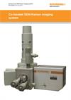
 Product note: Co-located SEM-Raman imaging system
Renishaw’s SEM-Raman system provides truly co-located analysis Renishaw’s SEM-Raman system is unique. You can simultaneously acquire both SEM (scanning electron microscope) and Raman data from the same area on the sample. You do not have to transfer the sample to a different measurement location or instrument; this ensures rapid truly correlative analysis. You will avoid the sample registration issues that can occur when moving samples between measurement locations.
[407kB] -
 Product note: SCA - diamond composite analysis
Renishaw’s structural and chemical analyser unites two well-established technologies, scanning electron microscopy (SEM) and Raman spectroscopy, resulting in a powerful new technique which allows morphological, elemental, chemical, physical, and electronic analysis without moving the sample between instruments.
[138kB] -
 Application note: A study of single-wall carbon nanotubes using Renishaw's structural and chemical analyser for scanning electron microscopy
Electron imaging and Raman spectroscopy are established techniques for viewing and analysing carbon nanotubes. Performing these two techniques usually requires the sample being transferred between a scanning electron microscope (SEM) and a Raman spectrometer. This application note illustrates the advantages of Renishaw’s structural and chemical analyser (SCA) for simultaneous secondary electron imaging and Raman spectroscopy of single-wall carbon nanotubes (SWNTs).
[357kB] -
 Application note: SCA oxidation application note - cultural heritage
SEM-SCA analysis helps in the preservation of a corroding bronze statue of the Roman god Ares from the ancient city of Zeugma in Turkey.
[354kB] -
 Application note: SCA oxidation application note - copper and steel
This note illustrates how the SEM-SCA can be used to study the corrosion of copper wire and nuclear reactor steel (four pages).
[345kB]
Downloads: Nanoindentation
-
 Application note: Indentation and Raman spectroscopy combined
Combine the power of inVia™ with nanoindentation measurements and directly correlate mechanical and tribological properties with chemical information such as crystallinity, polymorphism, phase and stress/strain. Raman spectroscopy is a powerful technique commonly used to study the composition, uniformity, strain, stress and disorder of materials. However, it does not directly characterise physical, mechanical and tribological properties. Renishaw and Hysitron have combined an inVia confocal Raman microscope with a TI 950 TriboIndenter, producing a system with the capability to directly correlate mechanical property measurements with comprehensive chemical analyses, in situ.
-
 News release: Combined Raman spectroscopy and nanoindentation assessment of complex materials
An article in the July/August 2016 issue of Microscopy and Analysis presents a new system that physically couples a Raman spectroscopy system with a nanoindenter on a shared stage that uniquely enables combined assessment of materials with submicrometre spatial resolution at single sites or spanning millimetre-sized regions.
Application examples
We have also produced a range of application examples, including the following.
If you would like to find out more, please contact your local representative using the button below, quoting the relevant document reference.
| Document reference | Document description |
| AS024 | AFM and 3D Raman imaging of glioma cells Reveal detailed and complementary information on the composition of glioma cells by atomic force microscopy (AFM) and inVia 3D Raman imaging. The AFM topograph—with nanometre-range spatial resolution—clearly identify cellular structures, but not their chemical compositions. By comparing and correlating AFM topographs with the additional chemical information from 3D Raman images, researchers can gain a better understanding of the cell. |
| AS027 | See sub-diffraction limit graphene features using tip enhanced Raman spectroscopy Reveal highly detailed graphene information by performing tip enhanced Raman spectroscopy (TERS) using inVia combined with an AFM. TERS uses a special plasmonic tip to increase the local electric field at the sample which, in turn, increases the Raman intensity. |
| AS028 | TERS of a malachite green monolayer Analyse minute sample volumes and weak Raman scatterers using inVia and TERS. Tip enhanced Raman spectroscopy (TERS) uses a special plasmonic tip to increase the electric field at the sample which, in turn, increases the Raman intensity. These tips are very small, with diameters on the order of 10 nm to 100 nm, and are held in contact with the sample using a scanning probe microscope (SPM) or atomic force microscope (AFM). |


