Deze pagina is momenteel niet beschikbaar in uw taal. U kunt met behulp van Google Translate een automatische vertaling bekijken. Wij zijn niet verantwoordelijk voor deze dienstverlening en het vertaalresultaat is niet door ons gecontroleerd.
Heeft u meer hulp nodig, neemt u dan contact met ons op.
Automatic focus tracking for Raman microscopy
LiveTrack™ focus-tracking technology automatically keeps the sample in focus on uneven surfaces. The microscope maintains focus on the sub-micrometre scale, even on surfaces that vary in height by millimetres. With LiveTrack technology, your Renishaw Raman microscope acts as an optical profilometer.
Raman microscopy on uneven surfaces
With LiveTrack technology, you can save time on sample preparation. You no longer need to prepare mineral sections or microtome pharmaceutical tablets. Raman analysis is easy even on large semiconductor wafers with a slight warp or bow.
LiveTrack technology is important for the most sensitive Raman measurements. This includes samples like nanoparticles or thin films. LiveTrack technology ensures that high numerical aperture (N.A.) objectives can stay in focus throughout data collection. The spectrometer thus collects the maximum Raman signal from the sample surface. Staying in focus also ensures that Raman images can have the highest spatial resolution possible.
The instrument uses continuous feedback to adjust the height of the sample stage. You simply focus on the sample and click once to activate LiveTrack technology. There is no need for any additional set-up time or pre-scans.
During microscopic inspection of uneven samples, LiveTrack technology removes the inconvenience of manually refocusing. WiRE™ software can also stitch together an in-focus video montage over large areas of an uneven sample.
Why use topographical Raman imaging?
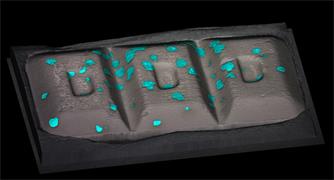
LiveTrack technology works with all of Renishaw's fast Raman imaging techniques. WiRE™ software can overlay Raman images onto 3D topographical views of the sample surface. The resulting 3D Raman mages can help you to understand how chemistry and structure relate to topography. You can also extract cross-sections of the samples in x- and y-planes for dimensional information.
Raman analysis during heating or cooling
Raman spectroscopy can perform in-situ studies on samples while varying conditions like temperature or humidity. If the sample surface moves in response to environmental factors, LiveTrack technology will maintain sharp focus. Your Renishaw Raman system can continue analysis on a sample in a temperature control stage. This is useful for studying the chemical or structural changes that occur during phase transitions or thermal annealing.

LiveTrack™ focus-tracking technology can maintain focus on a high-density polyethylene (HDPE) during melting and cooling. We placed a HDPE pellet in a crucible and heated it to 200 ˚C, before allowing it to cool to room temperature at a rate of 30 ˚C per minute. We recorded the Raman spectra and the z-position data.
Focus tracking for automated Raman measurements
LiveTrack focus-tracking technology is an essential part of Renishaw's fully automated Raman instruments. When you have activated LiveTrack technology, you can confidently stay in focus even when analysing multiple samples.
LiveTrack technology is available on the inVia™ confocal Raman microscope, the Virsa™ Raman analyser, the RA802 pharmaceutical analyser and the RA816 biological analyser.
Correlate surface topography with chemistry
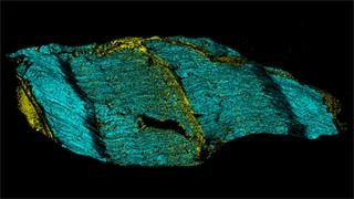
Understand the structure and chemistry of ammonite fossils.
Raman imaging of complex surfaces
Stay focused and always get high-quality Raman data. Activate LiveTrack technology on rough or dynamically changing samples.


