No momento esta página não está disponível em seu idioma. Você pode ver uma tradução automatizada utilizando o Google Translate. Não somos responsáveis pelo fornecimento deste serviço e não podemos verificar os resultados.
Se você necessitar mais ajuda fale conosco.
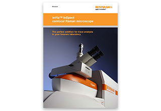
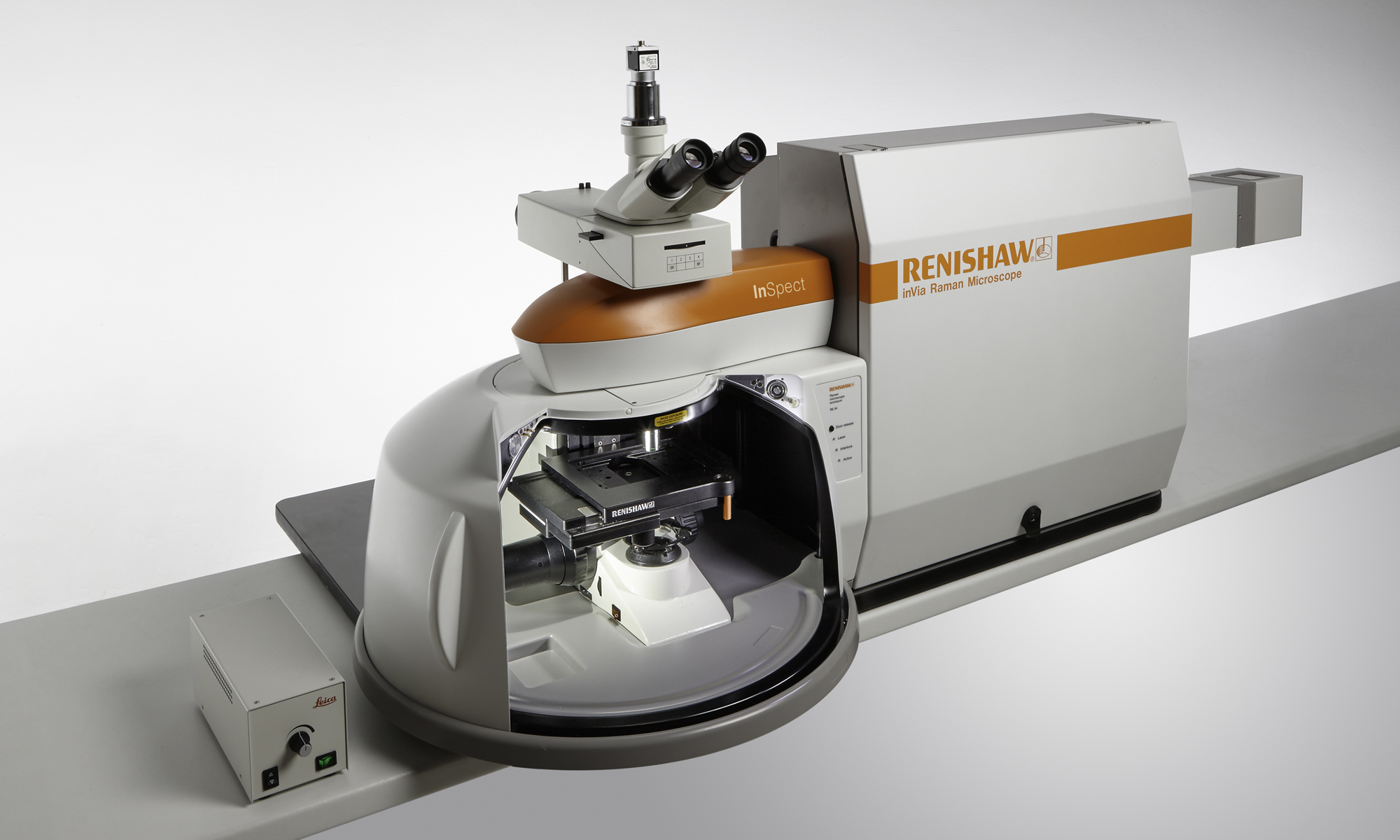
inVia™ InSpect confocal Raman microscope
Our best-selling confocal Raman microscope optimised for use in forensic laboratories for trace analysis.
Add Raman spectroscopy to your laboratory and gain further powerful capabilities which complement your existing techniques. Analysis is non-contacting and non-destructive and allows you to see fine chemical detail using a research-grade optical microscope.
Identify materials that may be difficult or time consuming with other techniques such as hard crystalline powders, ceramic shards and glass chips, easily analysed with little or no preparation required.
Features
Your inVia InSpect Raman microscope will give you:
- Highly specific identification - Raman microscopy can differentiate chemical structures, even closely related ones.
- High spatial resolution - comparable to your other microscopic techniques
- A range of microscope contrast techniques - including brightfield, darkfield and polarisation contrast with reflected and transmitted light illumination
- Particle analysis - use advanced image recognition algorithms and instrument control features to characterise particle distributions
- Correlative imaging - create composite images by combining Raman data with images from other microscopy techniques
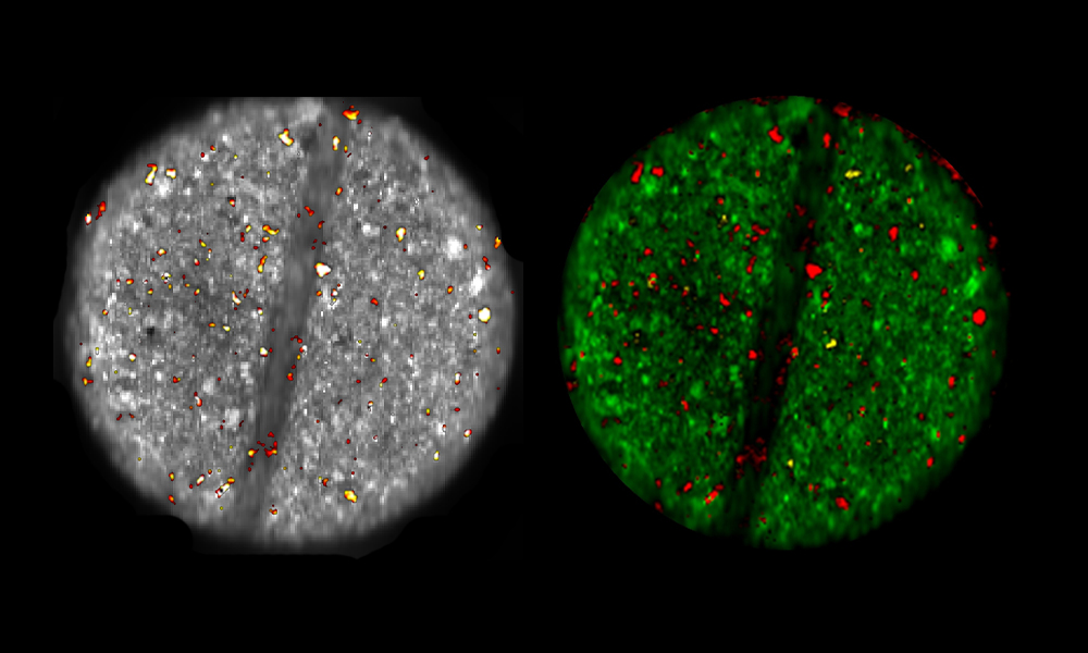
Empty Modelling™
Empty Modelling software uses a multivariate analysis technique to resolve complex data into its constituents. Use this, with library searching, to analyse data from samples that contain unknown materials.
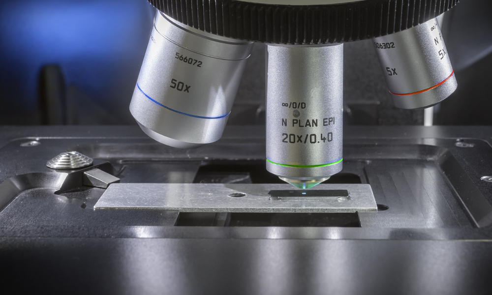
StreamHR™ Mapping
StreamHR mapping harmonises the operation of the InSpect microscopes high performance detector and MS30 microscope stage. It increases the speed of data collection and saves time generating images.
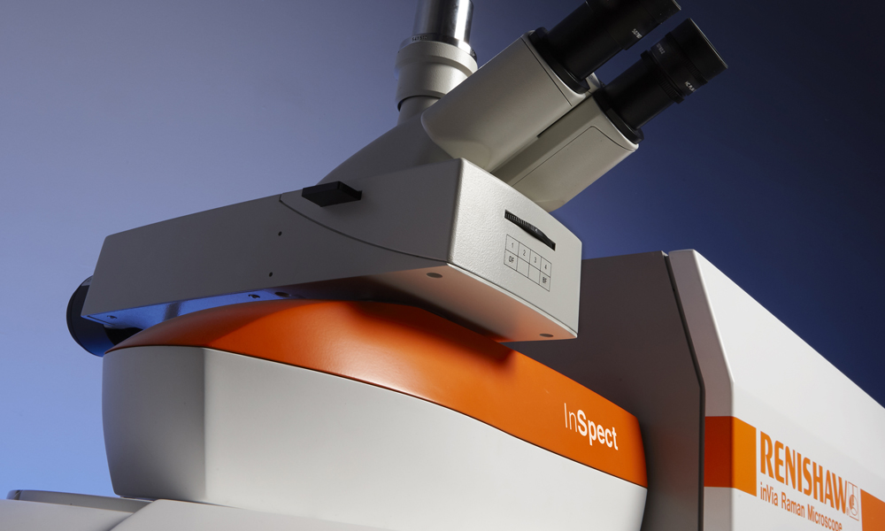
Full automation
You can control alignment, calibration, and configurations all through Renishaw's WiRE software. For example, you can rapidly switch—with a click of a button—between sample viewing and Raman analysis.
Applications
Gunshot residue
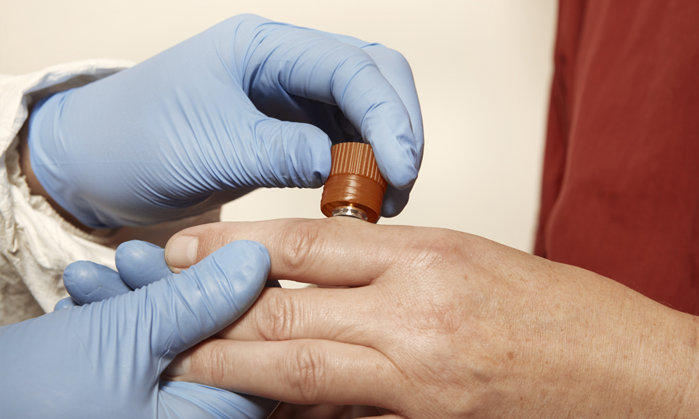
The inVia InSpect microscope provides a comprehensive package for the analysis of gunshot residue irrespective of its origin, delivering benefits for detection and identification of both organic and inorganic residues. In this note, we explore some of the key characteristics and benefits of the Raman technique for GSR analysis.
Document forgery
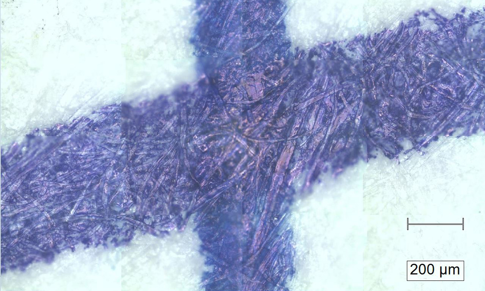
There are many different types of inks; colours may be the same, but chemically they can be different. Raman analysis using the inVia Raman microscope allows for rapid, non-destructive testing of questioned areas with the specificity to distinguish similar ink types that may visually look identical.
Want to find out more?
Your local representative will be happy to help with your enquiry.
You can contact them by completing a form or sending an email.
Get the latest news
Stay up-to date with our latest innovations, news, applications and product launches and receive updates directly to your inbox.
Specifications
Parameter | Value | |
| Wavelength range | 200 nm to 2200 nm | |
| Lasers supported | From 229 nm to 1064 nm | |
| Spectral resolution | 0.3 cm-1 (FWHM) | Highest typically necessary: 1 cm-1 |
| Stability | < ±0.01 cm-1 | Variation in the centre frequency of curve-fitted Si 520 cm-1 band, following repeat measurements. Achieved using a spectral resolution of 1 cm-1 or higher |
| Low wavenumber cut-off | 5 cm-1 | Lowest typically necessary: 100 cm-1 |
| High wavenumber cut-off | 30,000 cm-1 | Standard: 4,000 cm-1 |
| Spatial resolution (lateral) | 0.25 µm | Standard: 1 µm |
| Spatial resolution (axial) | < 1 µm | Standard: < 2 µm. Dependent on objective and laser |
| Detector size (standard) | 1024 pixel × 256 pixel | Other options available |
| Detector operating temperature | -70 °C | |
| Rayleigh filters supported | Unlimited | Up to four filter sets in automated mount. Unlimited additional filter sets supported by user-switchable accurately-locating kinematic mounts |
| Number of lasers supported | Unlimited | One as standard. Additional lasers beyond 4 require mounting on an optical table |
| Windows PC controlled | Latest specification Windows® PC | Includes PC workstation, monitor, keyboard and trackball |
| Supply voltage | 110 V AC to 240 V AC +10% -15% | |
| Supply frequency | 50 Hz or 60 Hz | |
| Typical power consumption (spectrometer) | 150 W | |
| Depth (dual-laser systems) | 930 mm | Dual laser baseplate |
| Depth (triple-laser systems) | 1116 mm | Triple laser baseplate |
| Depth (compact) | 610 mm | Up to three lasers (laser type dependant) |
| Typical mass (excluding lasers) | 90 kg |
