No momento esta página não está disponível em seu idioma. Você pode ver uma tradução automatizada utilizando o Google Translate. Não somos responsáveis pelo fornecimento deste serviço e não podemos verificar os resultados.
Se você necessitar mais ajuda fale conosco.
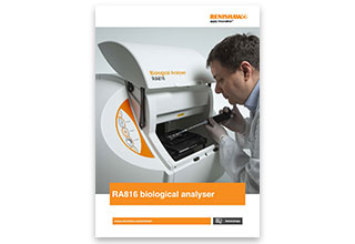
RA816 Biological Analyser
An easy to use and compact benchtop Raman imaging system that redefines
tissue and biofluid analysis.
The RA816 Biological Analyser enables you to identify and assess biochemical changes associated with disease formation and progression. There is little to no sample preparation, and no contrast agents or tags are needed.
Features
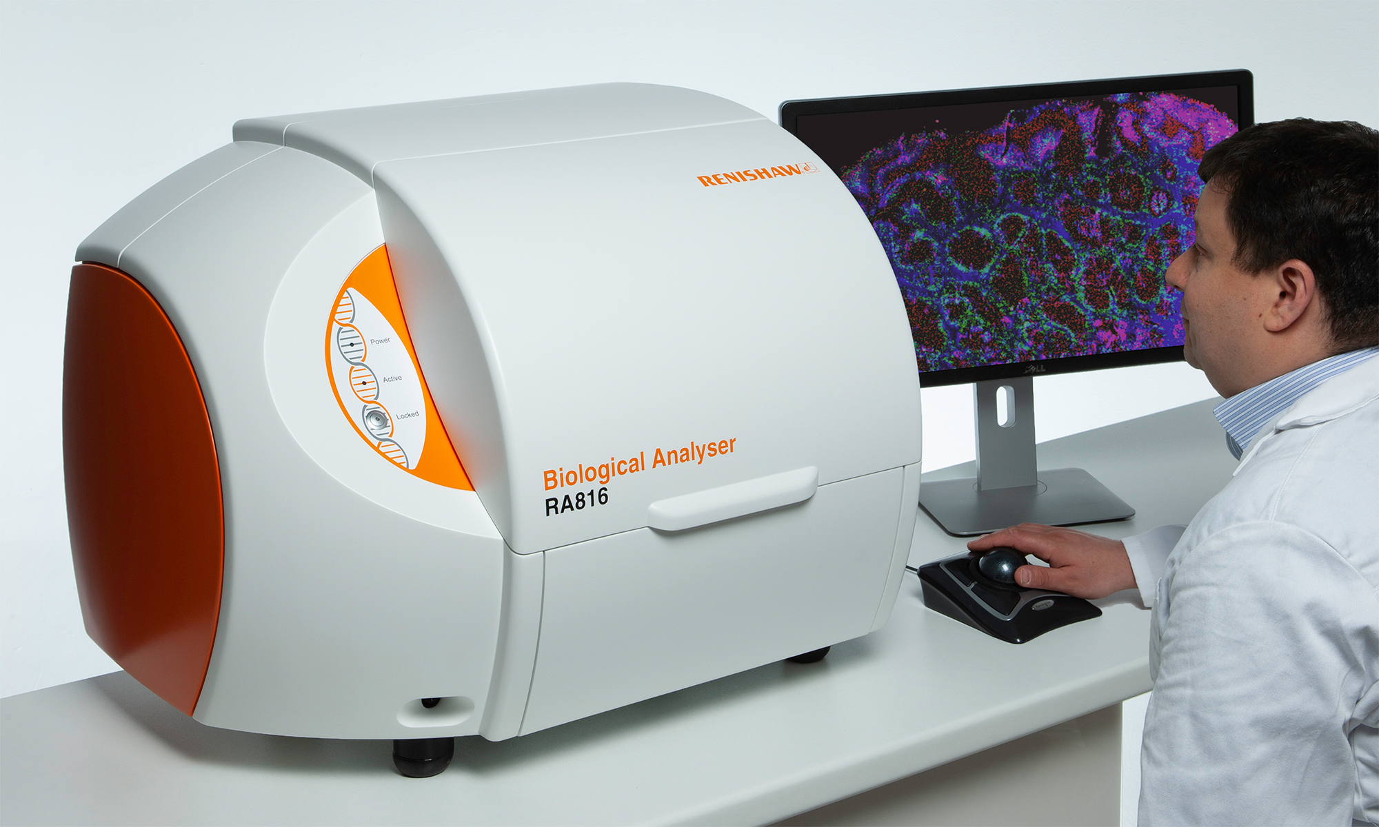
The RA816 Biological Analyser provides a practical solution for analysing biological samples. It is compact and transportable with optimised microscopic technology designed for biological and clinical sites.
- Easy to use hardware and software
- No need for stains or labels
- Minimal to no sample preparation
- Obtain full range of biochemical information (no need for prior knowledge of specific molecular targets)
Powerful software
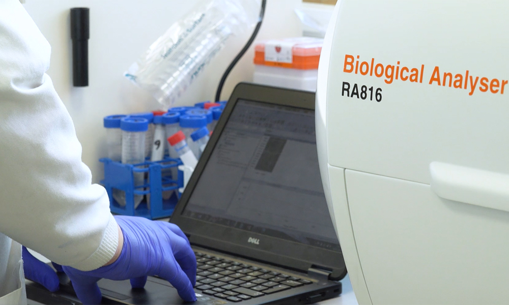
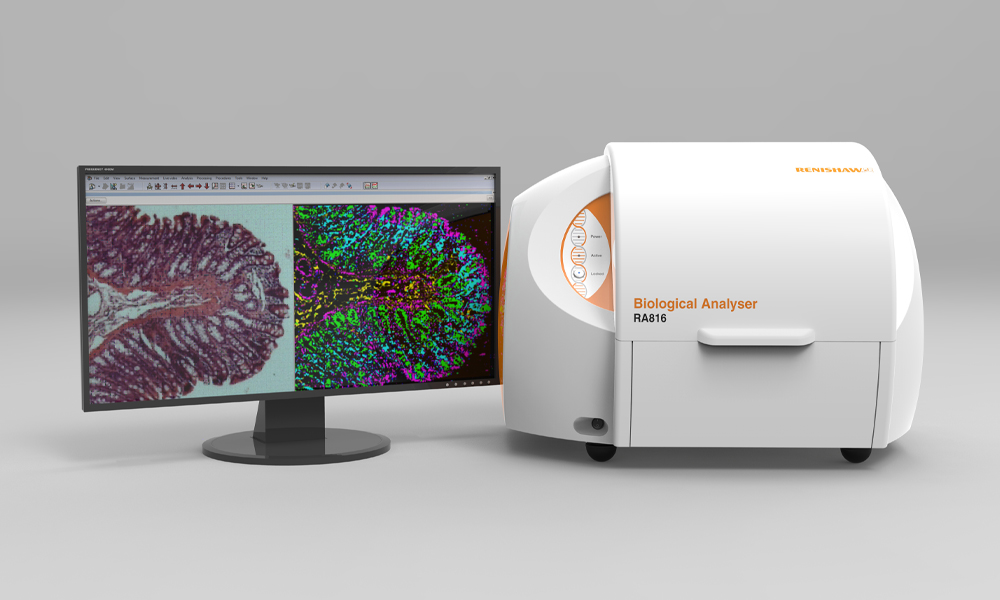
The RA816 Biological Analyser is entirely computer-controlled. Its software manages every step of the measurement process using pre-defined experiment setups. The unique macro image provides a comprehensive overview of all subsequent work and enables easy sample navigation and visualisation.
High performance
Stable and repeatable analysis with integrated performance qualification and alignment. It features LiveTrack™ technology which enables you to track sample surface and retain focus on challenging samples and Streamline™ technology for high speed data collection and image generation.
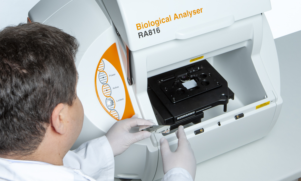
Easy to use
The RA816 Biological Analyser is ideal for the clinical research environment and can acquire data unattended. Its queuing capability enables you to configure measurements and leave the instrument to run them; you can analyse multiple samples on a slide without the need for user intervention.
Applications
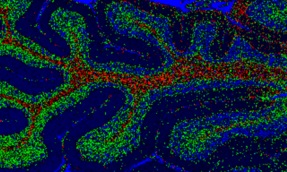
Classification of brain glioma tumours
In a study conducted with the University of Oxford Neuropathology Department in the UK, we demonstrated discrimination between diseased and healthy brain tissue using the RA816 Biological Analyser. We were able to differentiate diseased and healthy tissues without the need for disease marker discovery and targeting.
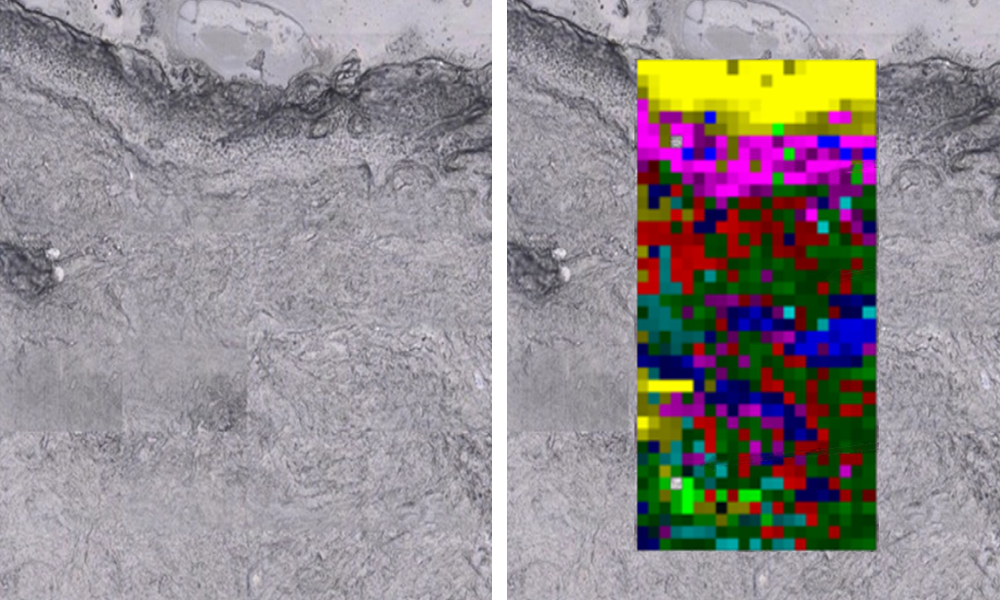
Monitoring transdermal drug delivery
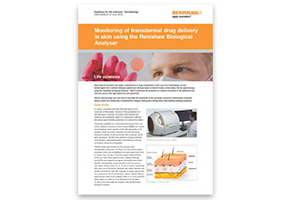
In a study, conducted with Miss Rubinder Basson from University of Manchester, Division of Musculoskeletal and Dermatological Sciences, the system used Empty modelling™ to demonstrate the presence and penetration depth of a compound in different skin tissue layers following application of a topical formulation.
Medical research using
Raman Spectroscopy
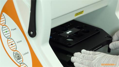
The Renishaw Biological Analyser brings together the chemical analysis power of Raman spectroscopy (a light scattering technique) and advanced optical and spectroscopic imaging technologies in a simple, robust system. It produces outstanding results, quickly and easily. Watch the video to find out more.
Want to find out more?
Your local representative will be happy to help with your enquiry.
You can contact them by completing a form or sending an email.
Get our latest updates
Stay up-to-date with our latest news, webinars, application notes and product launches delivered directly to your inbox.
Specifications
| Parameter | Value |
| Laser wavelength | 785 nm Integral Renishaw high power near infrared diode laser, 300 mW at 785 nm, air cooled, with integral plasma filter. Laser power: > 150 mW at sample. Innovative StreamLine technology enables higher laser power use without sample damage |
| Spectral range | 100 cm-1 to 3250 cm-1 Performed in two separate scan ranges Range 1: 100 cm-1 to 2000 cm-1; Range 2: 1950 cm-1 to 3250 cm-1 |
| Spectral dispersion | 2 cm-1 pixel-1 |
| Data collection speed | Over 1500 spectra/s |
| Minimum Raman image pixel size | 1 µm Spatial resolution 1 µm per pixel |
| Objective | 8.2 mm working distance 0.55 NA 50× long working distance objective Additional macro-view colour video camera |
| Field of view | Macro 21 mm × 16 mm Micro 330 µm × 250 µm |
| Maximum tiled image size | Macro 134 mm × 76 mm High magnification 112 mm × 81 mm |
| White light modes | White light transmission and reflection capability |
| Focusing | Macro – Manual or pre-defined Micro – Automatic (LiveTrack) or manual Real time automated LiveTrack dynamic focusing for both Raman data acquisition and white light video viewing modes |
| System calibration and transferability | Self-calibration and auto-align using built in neon and silicon references Automatic PQ data collection – (polystyrene) Optional post measurement check (PMC) for inter-measurement validation |
| Maximum sample size | ~ (110 mm × 90 mm × 25 mm) – fits 96 well plate |
| Power, voltage | 100 – 240 VAC ± 10%, 50/60 Hz, 100 W maximum |
| Dimensions | 720 mm (W) × 502 mm (H) × 535 mm (D) |
| Mass (not including computer) | 54 kg |
| Laser class | Class 1 laser product complies with IEC60825-1. CE marked |
