Por el momento, esta página no está disponible en español. Puede obtener una traducción automática mediante la opción de traducción de Google.
No podemos responsabilizarnos de este servicio puesto que podemos no verificar los resultados de la traducción.
Si desea más información, póngase en contacto con nosotros.
Feature articles
Get your research featured
Our feature articles contain blogs, opinions and research pieces from Renishaw applications scientists and our worldwide network of customers. If you use a Renishaw Raman system and have an article you'd like us to share, please get in touch.
Latest articles
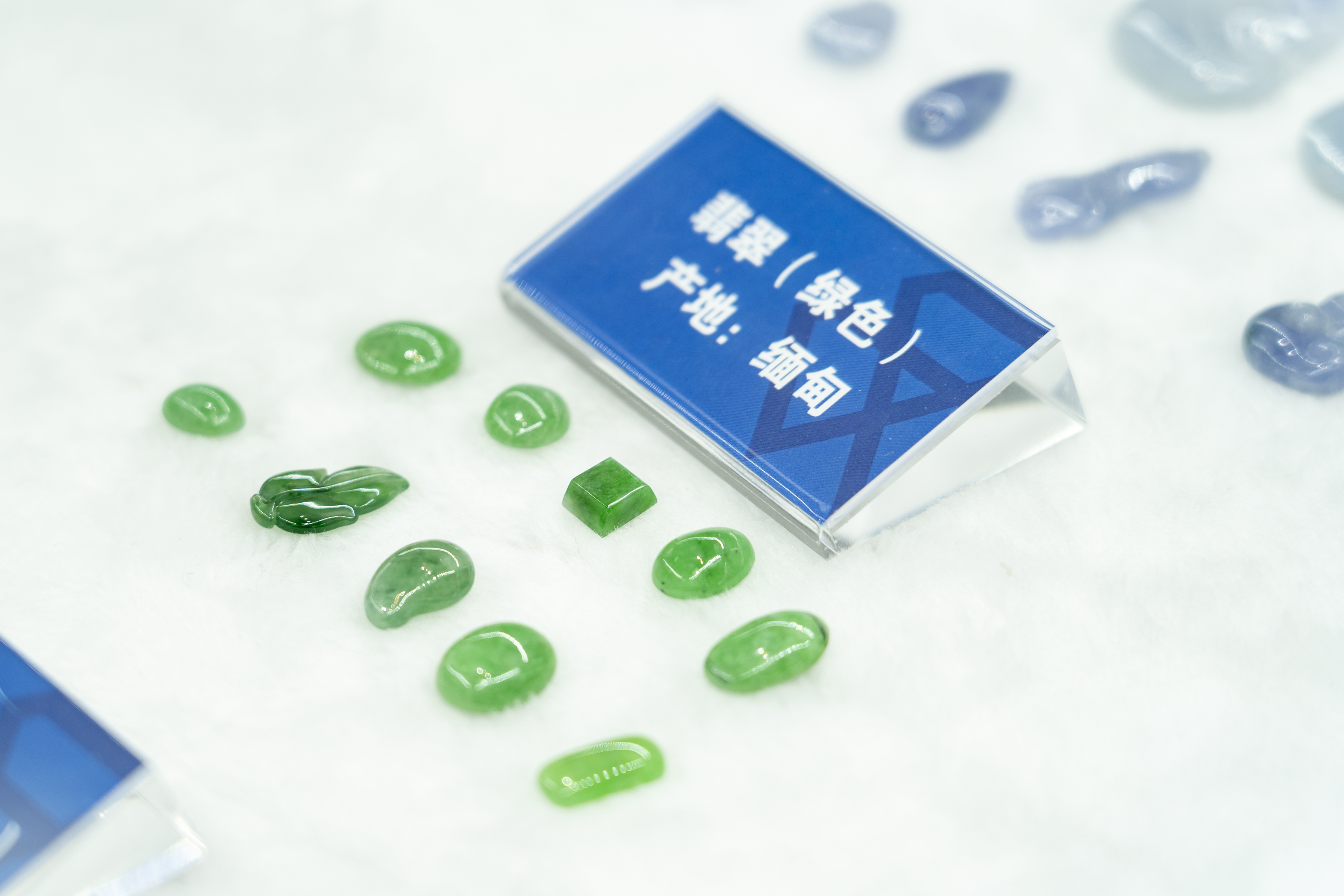
The Jewellery Institute of Guangzhou Panyu Polytechnic educates the latest generation of professionals for testing, appraisal, assessment and standards development in the jewellery industry. With the increasing demand for instrument technology at the school, confocal Raman microscopes offer enormous advantages for daily teaching and analytical testing across a wide range of applications.
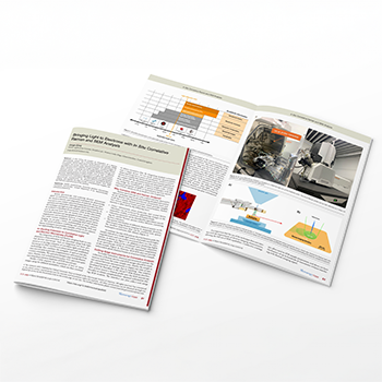
Our Applications Scientist, Jorge Diniz, has authored an article for the July 2025 issue of Microscopy Today. He describes Renishaw’s integration of a Raman spectroscopy system with a scanning electron microscope (SEM). Using examples such as a battery electrode, a geological section, and contaminants on a fuel injector, Jorge explains how correlative SEM and Raman analysis can provide a greater understanding of the chemistry and structure of samples.

Dr. Yin Xia, the Director of the Relics Conservation and Restoration Department at the Emperor Qin Shihuang's Mausoleum Site Museum, has used an inVia™ confocal Raman microscope to help identify pigments on the terracotta figures for their conservation and restoration. To commemorate 50 years since the discovery of the Qin Terracotta Warriors, we asked Dr Xia to share some his insights into the conservation of these ancient relics.

Scientists in France have been studying charred material from archaeological sites with Raman spectroscopy. They showed that it is possible to determine what material was charred and at what temperatures. After the fire at Notre Dame cathedral in Paris, France in 2019, the researchers applied this method to determine the highest temperatures reached in the roof structure. In addition, they analysed chars from the Bruniquel caves in Southern France to show either animal or vegetal precursors.

Located in Salisbury, UK, Stonehenge is the most architecturally sophisticated prehistoric stone circle in the world. In the quest to unravel the mysteries of Stonehenge, Renishaw had the privilege to assist in analysing the Altar Stone in situ using a Renishaw Virsa™ Raman analyser. The Altar Stone is unusual and differs in appearance when compared to the other main bluestones of Stonehenge. The aim was to use Raman spectroscopy and minerology to understand the provenance of the stone.

We show how Raman spectroscopy is being used to investigate nanostructured materials for carbon capture. With climate change and rising emissions, governments are looking at new ways to reduce atmospheric carbon dioxide concentrations we show how Raman spectroscopy is being used to investigate nanostructured materials for carbon capture. With climate change and rising carbon dioxide emissions, governments are looking at new ways to reduce atmospheric concentrations.
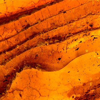
In recent years, Renishaw Raman microscopes, located at two prestigious geological institutions, have been used for the non-destructive analysis of inclusions in ultradeep diamonds from the Earth’s lower mantle. Diamond inclusions are an important chemically inert vessel, preserving the original structure and chemistry of mineral fragments formed under high pressure.
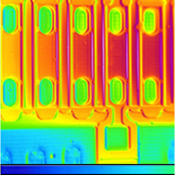
Thermography microscopes can efficiently map high-temperature zones over large surface areas, but the challenge is being able to pinpoint minute areas of high heat. To address this, Quantum Focus Instruments (QFI) coupled a Renishaw Virsa Raman analyser to their InfraScope, enabling interesting Raman thermographic findings.
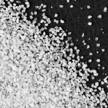
At the atomic-scale, glass structure is typical of a supercooled liquid. However, glass simultaneously has all the mechanical properties of a solid on a macroscopic scale. Raman spectroscopy is a powerful technique for analysing glass. It gives chemical and structural information, is non-destructive, and does not require any sample preparation.
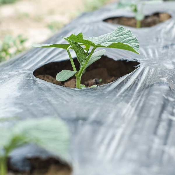
Crop producers commonly use plastic mulch films to suppress weeds, conserve water, and prevent contamination from atmospheric agents. However, they are difficult to recycle and there is a risk of microplastics derived from mulch film (MMF) polluting the wider environment. Researchers at Guangxi University, China, and the Chinese Academy of Tropical Agricultural Sciences have used Raman spectroscopy to determine the portion of microplastics which derive from agricultural mulch films.
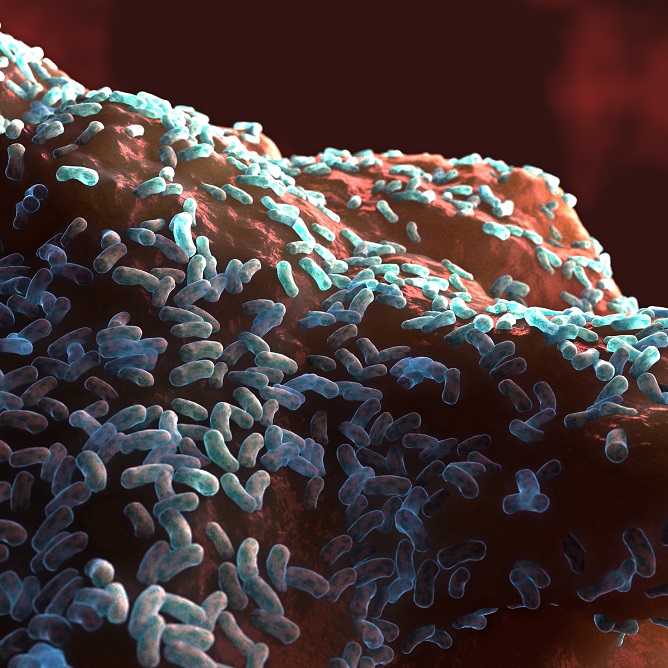
We summarise a study carried out by Proctor & Gamble and by the Fraunhofer Institute for Interfacial Engineering and Biotechnology which used a Renishaw inVia confocal Raman microscope and a multivariate analysis to spatially differentiate bacteria in an in vitro model simulating a subgingival dual-species biofilm.

Renishaw is featured in the Jan/Feb 2022 issue of Microscopy and Analysis magazine. Renishaw’s head of spectroscopy applications Tim Smith has written an article on the importance of software functionality for a modern Raman spectrometer. In this article, Tim describes how software for Raman spectroscopy systems has advanced over recent years and how it is a valuable asset to your Raman system.

The Raman technique is ideal for analysing cultural heritage pieces as the ability to examine analyse objects in situ using the sizeable chamber of the Renishaw inVia confocal Raman microscope meant that sampling could be done non-destructively.
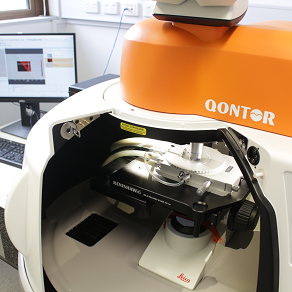
The Renishaw inVia confocal Raman microscope was chosen as the spectrometer of choice for the MCFP, as its bespoke configuration is perfectly suited to analysing cultural artefacts with varying topography and composition.
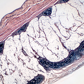
A group of researchers and clinicians at University College London, Royal Veterinary College and Charing Cross Hospital, led by Dr Riana Gaifulina, have demonstrated that Raman spectroscopy could determine the grade of cartilage erosion and overall osteoarthritis disease state. This could lead to an earlier identification and treatment of osteoarthritic degeneration.
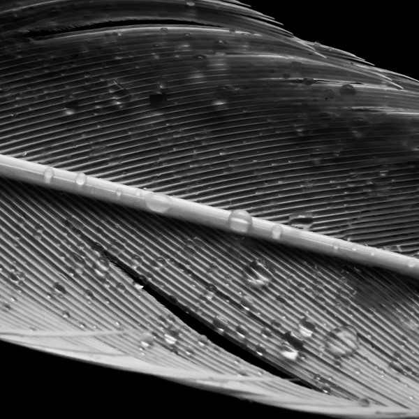
Raman technique was chosen as the data collection method for this PhD thesis as it provides a high-precision analysis of the proteins contained within flight feathers to elucidate their secondary structures.

How can a surgeon accurately distinguish tumour tissue from healthy tissue in under 15 minutes? The answer could be to use Raman spectroscopy.
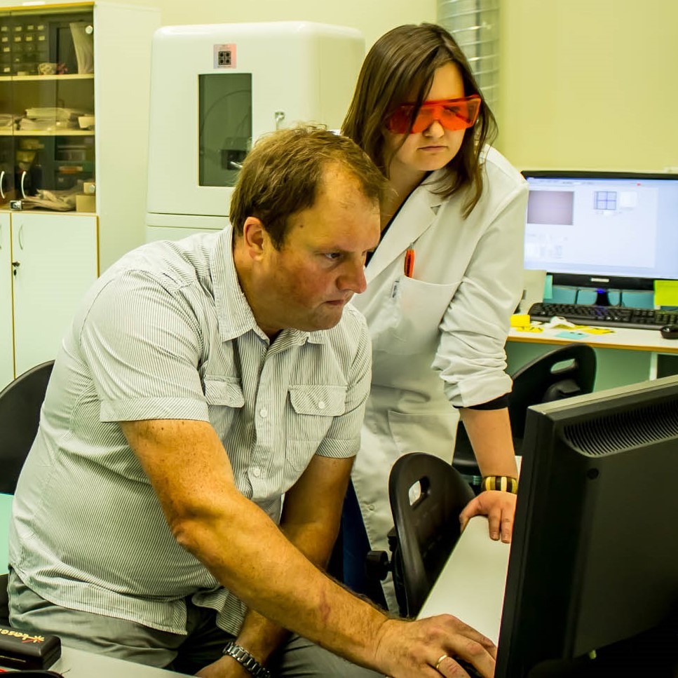
New Plasma Technologies uses a Renishaw inVia confocal Raman microscope to control the quality of the coatings and help improve processing methods, discover why.
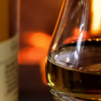
Whiskies are big business, so having an objective, scientifically robust, method to evidence the authenticity of the product is important to ensure counterfeits or diluted/mixed products are identified. We show how the inVia Raman microscope has been used to analyse a variety of whiskies through their unopened bottles to evidence their authenticity.

It is vital that we monitor plants and detect changes taking place, ideally before the visible signs of them occur. We therefore need suitable analytical techniques. Raman spectroscopy is ideal for this.

A team of researchers from the CHUM Research Centre and the Polytechnique Montreal have developed a new technique to improve detection of aggressive forms of prostate cancer.
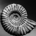
A sensitive Raman system with a high spectral resolution is essential for the successful analysis of geological samples. We put StreamLine Raman imaging to the test by analysing an ammonite fossil.

Sun lotion is a highly advanced emulsion, a true example of human endeavour against nature, and ultimately a life saver. So let’s look deeper into this powerful cream using the inVia™ confocal Raman microscope.

Raman analysis allows for rapid, non-destructive testing of questioned areas with the specificity to distinguish similar ink types that may visually look identical. We test a method for analysing crossing ink lines to identify possible forgeries.
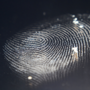
To be truly useful, in a highly practical and physical environment, chemical imaging needs to be versatile and forgiving over a range of object surfaces. We put Raman imaging to the test by analysing a fingerprint on the side of a soda can.
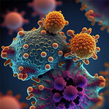
Researchers used a Renishaw RA816 Biological Analyser to identify the genetically different subtypes of brain gliomas in a neurosurgical setting. By understanding the genetics of a tumour during the operation, the clinician would be able to map out the best surgical and treatment strategy. This could improve survival rates and treatment response of the patient.

Utilising a Renishaw Raman spectroscopy system, a team of researchers from the Czech Academy of Sciences have been testing a novel way to identify Staphylococcal bacteria, paving the way for faster diagnosis and treatment of infectious diseases.
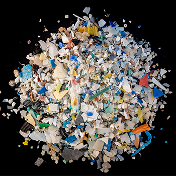
Could rubber and plastic from tyres, washing into rivers and streams, be contributing to pollution in waste water? A group of scientists from Denmark are using a system from Renishaw to find out.

In January 2017, the world’s lightest mechanical chronograph watch was unveiled in Geneva, Switzerland, showcasing innovative composite development by using graphene. Now the research behind the project has been published.