CCD detectors for Raman spectroscopy
Renishaw's Raman instruments come equipped with the Centrus™ detector. We have designed the Centrus detector with high speed and high sensitivity for both Raman and photoluminescence (PL) spectroscopy. Our high-performance Centrus detector has excellent quantum efficiency across a broad spectral range, from the ultraviolet (UV) to the near-infrared (NIR) wavelengths. The Centrus detector enables fast chemical detection and imaging on all of Renishaw's Raman spectroscopy systems.
Highly sensitive spectroscopy with a low-noise CCD detector
When we equip a high throughput inVia™ confocal Raman microscope with the Centrus detector, it is capable of the most challenging spectroscopic measurements. The Centrus detector is a thermoelectrically cooled charge-coupled device (CCD) detector. It has a high quantum efficiency and ultra-low readout noise to detect even the weakest Raman bands.
If required, we can configure an inVia microscope with up to 4 different detectors. This includes back-illuminated (BI) or electron-multiplying (EM) CCD detectors. Get in touch to find out if these options are suitable for your sample type.
Ultra-fast detector readout for Raman imaging
With the Centrus detector, you can take advantage of Renishaw's ultra-fast Raman imaging techniques. You can also run rapid time-series measurements to analyse chemical reactions. Using Renishaw's patented electronic designs, the Centrus detector can achieve a high-speed readout rate of 2 MHz. This enables the collection of full-range, high-resolution spectra at rates faster than 1800 spectra per second.
Raman spectroscopy from UV to IR
With a single Centrus detector, you can collect high-quality Raman and PL spectra with laser excitations ranging from 229 nm to 830 nm. Our CCD detector has a high detection efficiency across a broad range of wavelengths, from the deep-ultraviolet (DUV) to the near-infrared (NIR). In combination with the inVia microscope's full automation and kinematically-mounted spectrometer optics, you can easily use a wide spectral range.
For coloured samples with high fluorescence backgrounds in the visible range, Raman spectroscopy using an excitation laser at 1064 nm can help to reveal Raman bands. Choose an inVia microscope with an InGaAs detector for Raman spectroscopy at longer infrared wavelengths.

Broadband spectrum of SiC showing photoluminescence (PL) features. The spectral range is from 400 nm to beyond 1000 nm. This spectral range is over 10,000 cm-1, from the visible blue wavelengths to beyond the infrared. Data courtesy of Prof. John Steeds and Dr. Geraint Evans, Dept of Physics, University of Bristol, UK.
Extended scanning for a complete Raman spectrum
Centrus detectors support SynchroScan™ extended scanning technology. With SynchroScan technology, you can easily collect artefact-free continuous spectra over a broad spectral range. SynchroScan technology does not compromise on high spectral resolution. You do not have to use a diffraction grating with lower dispersion to extend the spectral range.
Extended scanning is particularly useful when:
• Identifying similar materials such as polymers or pharmaceutical polymorphs.
For reliable identification, extended Raman spectra should contain the Raman bands in the fingerprint region (<1500 cm-1). Raman spectra should also include the CH2 and CH3 stretching modes (between 2800 cm-1 and 3300 cm-1).
• Resolving both broad and sharp features in the PL spectra from semiconductor materials like SiC.
Scientists use PL spectra to study the electronic properties and the presence of defects in semiconductors. This often requires a spectral range of hundreds of nanometers (or thousands of wavenumbers, cm-1). For an inVia™ confocal Raman microscope with a 532 nm laser, SynchroScan technology can cover a spectral range of up to 10,000 cm-1 in a single extended scan.
SynchroScan technology uses a patented method to synchronise the stepwise rotation of the diffraction grating with continuous readout at the detector. As the diffraction grating rotates, the light on the detector moves in the direction of linear dispersion. To keep in step with this movement, the signal also moves along to the next pixel. When the accumulated signal gets to the edge of the Centrus detector, the software records the next point on the spectrum. For further details see C. Dyer & B.J.E. Smith, Journal of Raman Spectroscopy, Vol. 26, 777-783 (1995).
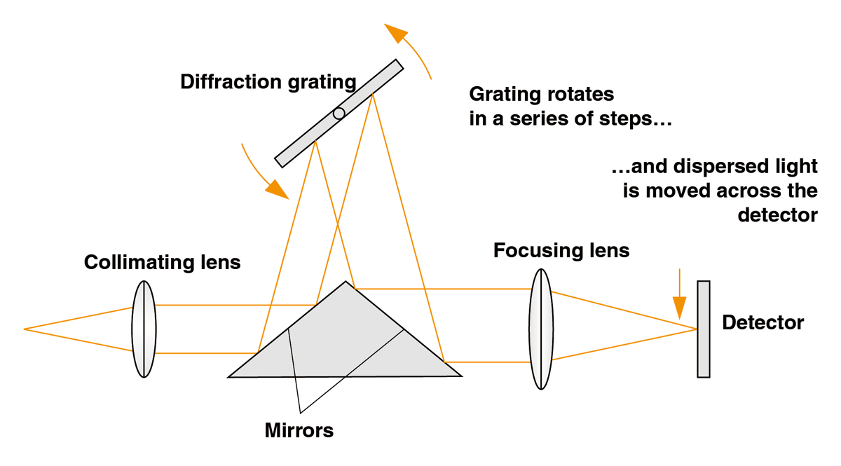 Schematic showing the synchronised movement of the diffraction grating and signal across the detector.
Download document
Many aspects of Renishaw's innovative technology are covered by patents.