Multiple lasers and gratings for Raman and PL spectroscopy
To give you full flexibility when analysing with different laser wavelengths and spectral ranges, key optical components within the inVia™ confocal Raman microscope can be easily swapped. These include parts such as diffraction gratings, filters and spectrometer lenses. Renishaw's encoded stages and kinematic mounts ensure accurate repositioning of spectrometer optics. This saves time by avoiding manual realignment.
Ultraviolet, visible and infrared lasers for Raman spectroscopy
To perform Raman spectroscopy, we focus a single colour of light onto a sample with and analyse the resulting Raman-scattered light. Lasers are the ideal light source for Raman spectroscopy as they emit intense, highly collimated and monochromatic light. The excitation laser wavelength affects the resulting Raman intensity, spatial resolution and fluorescence background. It is important to choose suitable laser wavelengths for your Raman instrument.
The inVia™ confocal Raman microscope offers the widest range of excitation laser options, including:
• Ultraviolet (UV) laser wavelengths: 229 nm, 244 nm, 266 nm, 325 nm, 355 nm, 364 nm.
• Visible laser wavelengths: 405 nm, 442 nm, 457 nm, 473 nm, 488 nm, 514 nm, 532 nm, 633 nm, 647 nm, 660 nm.
• Infrared (IR) laser wavelengths: 785 nm, 830 nm, 1064 nm.
If the sample contains a fluorophore, Raman analysis with visible lasers can lead to spectra containing strong fluorescence backgrounds. These high backgrounds can obscure the Raman bands. Switching to near-IR or UV lasers can help to avoid fluorescence. This approach is often useful for analysing polymers and organic materials.
For certain samples, the use of UV excitation lasers can take advantage of resonance phenomena to boost Raman signals significantly. Resonance can enhance the sensitivity of a Raman measurement and thus enable the detection of thin molecular films.
Durable kinematic mounts
The contact surfaces of Renishaw's kinematic mounts are tungsten carbide. This material is very hard and is more wear-resistant than steel. This ensures a lifetime of reliable and stable operation from the mounts.
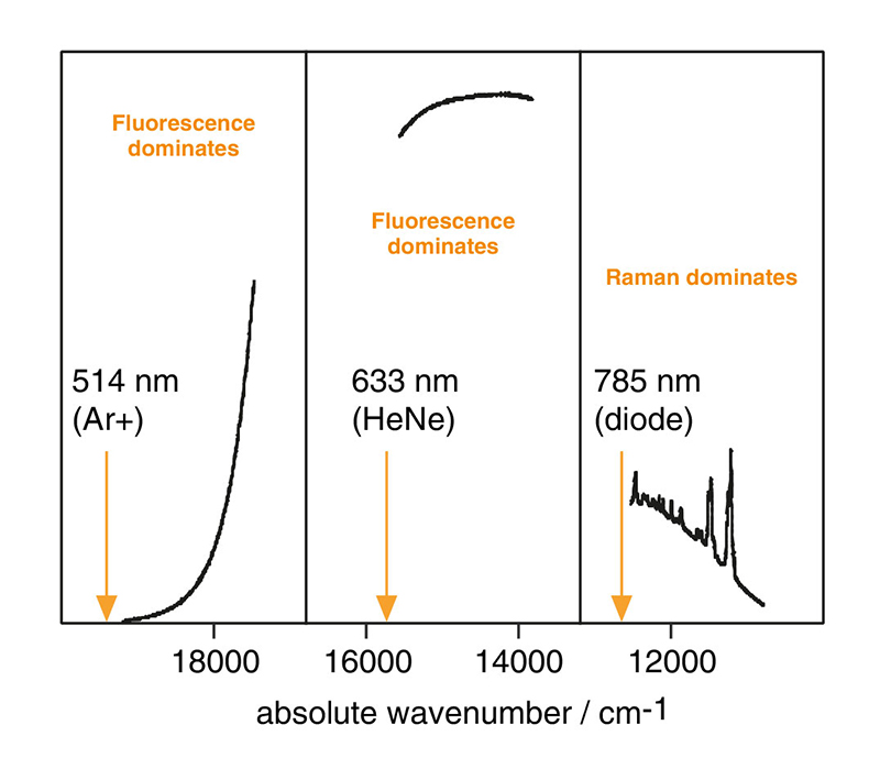 Spectra from a conducting polymer using different laser wavelengths. The near-IR 785 nm spectrum clearly shows Raman bands. In contrast, the visible spectra using either 514 nm or 633 nm contain strong fluorescence backgrounds.
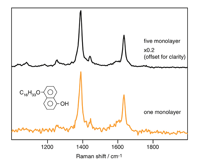
UV-resonance Raman spectra of molecular monolayers. Data courtesy of Dr. Zhou Bing, Institute of Theoretical Chemistry, Jilin University, Peoples' Republic of China.
What makes a good Raman spectrometer?
This webinar series explores Raman spectrometer configurations including lasers, lenses, gratings and detectors. Learn how these choices can enhance sensitivity, spatial resolution and spectral resolution.
Rayleigh-rejection filters for Raman analysis
During Raman analysis, most of the light that falls on a sample is elastically scattered. This Rayleigh-scattered light is typically 10 million times stronger than the Raman-scattered light. To successfully perform Raman analysis, we use Rayleigh-rejection filters to stop the elastically scattered light from entering the spectrometer. Typically, each laser wavelength requires one Rayleigh filter set. Most users do not manually change filters because the inVia microscope can automatically swap between 4 Rayleigh filter sets.
A range of Rayleigh filter technologies are compatible with the inVia microscope. Our most popular Rayleigh filters are high-efficiency edge filters for detecting Raman bands down to 100 cm-1. You can also configure an inVia microscope with:
• ripple-free, broadband filters for photoluminescence (PL) spectroscopy.
• notch filters for observing both Stokes and anti-Stokes Raman bands.
• low-cutoff filters for low-frequency Raman analysis.
Filters for low-frequency Raman analysis
An inVia microscope supports a range of low-wavenumber, or low-frequency filter options. Our range of Eclipse filters can analyse Raman bands down to ca. 10 cm-1. These enable you to study samples with low-energy conformational changes, crystalline lattice modes, vibrational modes involving heavy atoms, and the rotational behaviour of gases.
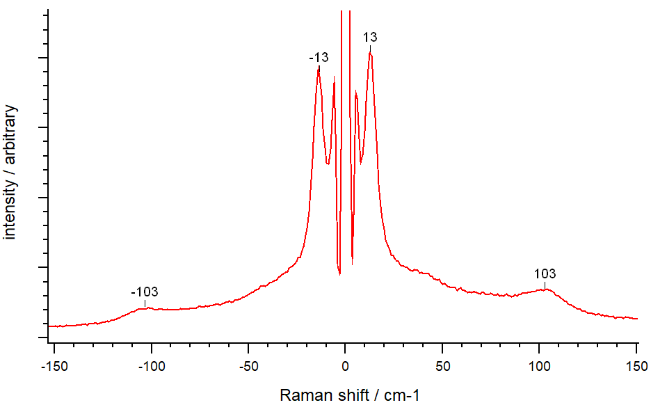 Low-frequency Raman spectrum showing the longitudinal acoustic mode (LAM) of high-density polyethylene (HDPE), for both the Stokes and anti-Stokes bands at ~13 cm-1. Renishaw's range of Eclipse filters can analyse Raman bands down to ~10 cm-1.
Swap spectrometer optics easily with kinematic mounts
On an inVia microscope, you can easily swap between a full range of optics using Renishaw's kinematic mounts. Kinematic mounts ensure accurate and repeatable positioning of filters, lenses and gratings. This gives you full flexibility without any need to manually realign. For example, Raman analysis with either a UV or 1064 nm infrared laser often requires different lenses and diffraction gratings. You can easily swap these optics in less than 5 minutes.
The contact surfaces of our patented kinematic mounts are made of tungsten carbide. This material is very hard and is more wear-resistant than steel. This ensures that optics in a Renishaw spectrometer are always in the right place.
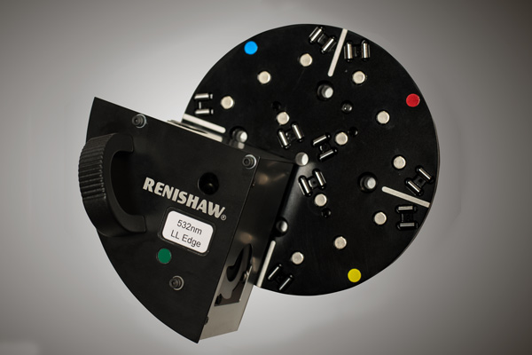
Renishaw's kinematic mount for Rayleigh filters within the inVia™ confocal Raman microscope.
Does my Raman spectrometer need more diffraction gratings?
An inVia microscope can automatically swap between two diffraction gratings. There is often no need to manually exchange any optical components. In addition, SynchroScan™ extended scanning technology can collect high-resolution spectra across a wide spectral range without changing the diffraction grating.
Renishaw's Raman systems use diffraction gratings to separate the Raman-scattered light into its constituent wavelengths. Within the spectrometer, a lens focuses the dispersed light onto the pixels of the CCD detector so it can be read off as a Raman spectrum. Typically, each laser wavelength is paired with one diffraction grating. On the inVia microscope, we usually use different diffraction gratings with UV, visible and IR excitation lasers in order to achieve a spectral resolution of ca. 1 cm-1. A Raman spectral resolution of 1 cm-1 is more than enough to analyse most samples.
On an inVia microscope, you can easily swap between more diffraction gratings using Renishaw's kinematics mounts. Choose a diffraction grating with a higher groove density to achieve a high spectral resolution of 0.3 cm-1. This could help to resolve very narrow Raman bands from highly crystalline solids, or the rotational modes of gases. Conversely, for photoluminescence (PL) spectroscopy across abroad spectral range, our spectrometers can be set up with gratings that have a lower groove density.
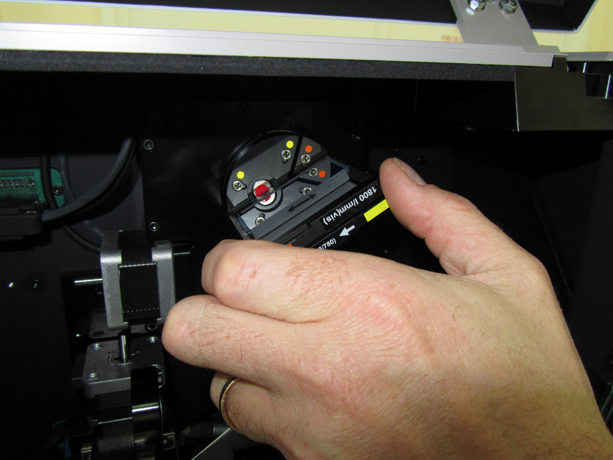
A user changes diffraction gratings using kinematic mounts in an inViaTM confocal Raman microscope.
Download document