High-speed Raman and PL imaging
You can perform Raman spectral imaging to show the 2D and 3D spatial distribution of chemical species within a sample. As a result, you can better understand the chemical components within your sample. Most samples are not homogeneous; you need to know the size and distribution of their components to understand them fully. You can also determine the structural variation in a material, such as crystallinity, stress state or number of layers.
Efficient Raman imaging in less time
StreamHR™ and StreamLine™ imaging technologies can analyse your sample with either Raman or PL imaging in the shortest possible time. With data collection rates up to 1,500 spectra per second, you can get Raman images in minutes. Both methods save time by streaming spectral data from the CCD detector while scanning the excitation laser over the sample.
Renishaw's fast imaging methods can obtain chemical images from large sample areas. The analysed area can be larger than the field of view of the microscope objective. Raman instruments with an MS30 high speed encoded stage can analyse sample areas as large as 112 mm × 76 mm. For high-speed imaging on even larger samples, we can configure an inVia microscope as a free-space microscope.
StreamHR™ and StreamLine™ rapid imaging technologies:
• For Raman or photoluminescence (PL) imaging
• Save time with continuous spectral data readout
• Sample areas up to 112 mm × 76 mm with an MS30 stage
• Use on uneven samples with LiveTrack™ focus-tracking technology
• Configure multiple Raman or PL images with batch measurements
With both StreamHR and StreamLine technologies, LiveTrack™ focus-tracking technology can automatically maintain focus on uneven samples in real time. You can also automate the Raman imaging of multiple positions using queuing or batch measurements.
StreamHR™ imaging technology: high-resolution Raman imaging
If your sample contains features smaller than 1 µm, you might need the highest spatial resolution to reveal them. StreamHR technology scans the sample with a diffraction-limited laser spot at small step sizes, down to 50 nm. This produces chemical and structural information as 1D profiles, 2D areas or 3D volumes with the highest possible detail.
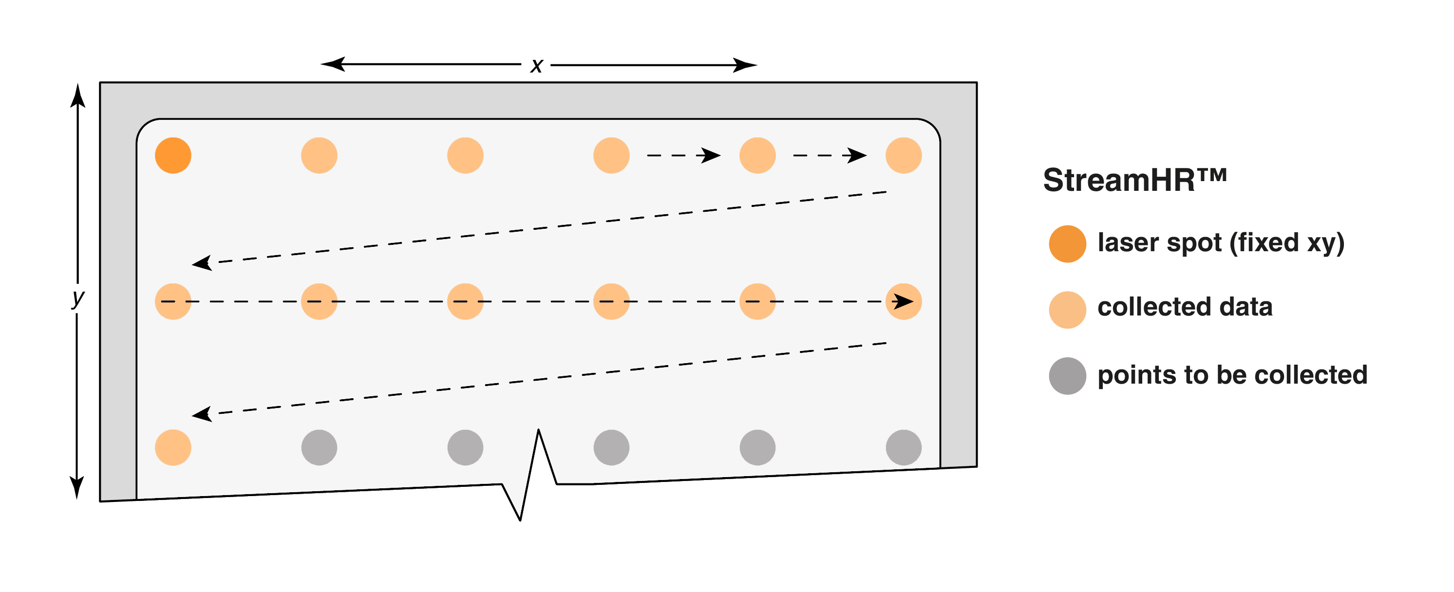
Data collection using StreamHRTM imaging technology. The sample moves under the laser spot while the CCD detector continuously reads spectral data. The step size between pixels can be as low as 50 nm.
StreamLine™ imaging technology: Fast and non-destructive Raman imaging
StreamLine technology rapidly generates chemical images of large sample areas. It spreads the laser power into a line of laser light, rather than an intense spot. This prevents laser-induced sample damage and efficiently samples an entire line of points at the same time.
StreamLine technology scans the laser line across the sample in time with the movement of the signal on the detector. Each imaged point of the sample traverses the laser line once. As it does so, the detector accumulates the signal and then reads it. This method can rapidly collect large, artefact-free chemical images with XY spatial resolution around 1 µm.
Image gallery


Even faster Raman imaging in Rapide mode
If you need even faster Raman imaging, use StreamHR or StreamLine technology in Rapide mode for that extra boost. The Rapide option uses a patented method to synchronise sample stage and the CCD detector readout at extreme speeds.
StreamHR™ Rapide imaging technology can collect highly resolved Raman images at rates faster than 1,000 spectra per second. You can also use the ultra-fast spectral readout of StreamHR Rapide technology to acquire data from the same point. This is useful for monitoring fast chemical reactions and processes down to microseconds.
For the fastest Raman imaging, StreamLine™ Rapide imaging technology can collect data at rates faster than 1,500 spectra per second. This method is suitable for Raman imaging of larger areas at the highest speeds.
StreamHR™ Rapide and StreamLine™ Rapide imaging technologies are available on the inVia™ confocal Raman microscope.
Raman images explained
Want to learn more about Raman images? Get started quickly with the fundamentals of Raman imaging.
FAQ
1. What is the difference between Raman imaging and Raman mapping? Are they the same thing?
In the past, ‘Raman mapping' referred to the process of collecting Raman spectra from each position on a sample. The collected data is a spectral hypercube that can be analysed to produce Raman images.
Currently, published scientific literature uses both ‘Raman imaging' and ‘Raman mapping' interchangeably. Most researchers will understand Raman imaging and Raman mapping to be the same thing.
2. Why should I use binning when collecting spectral images with StreamLine technology?
StreamLine technology is ideal for fast chemical imaging domains (or particles) down to 1 µm in size. If you do not require high spatial resolution, you can choose to enhance the signal-to-noise ratio and the data collection speed by ‘binning.' If you use binning, this adds together the spectral data from a specified number of rows on the CCD detector. This drastically reduces the exposure time and the total data collection time. For more information, please read (PN on fast Raman imaging).
3. Why is the Slalom mode useful for StreamLine technology?
You can use StreamLine technology with Slalom mode to solve the problem of undersampling. If you use step (pixel) sizes that are larger than the width of the laser spot or line, Raman imaging might not detect smaller particles.
With Slalom mode, the laser line moves in a zig-zag motion to collect data from the entire region of interest. This helps to detect smaller features that you could have missed. Such small features could be monolayer graphene on a large wafer, forensic contaminants, semiconductor defects or SERS nanoparticles. For more information, please read (PN on fast Raman imaging)
Download document
Many aspects of Renishaw's innovative technology are covered by patents.